


Veterinary microscope: how to choose and use it in your clinic
A guide to choosing a Euromex bScope veterinary microscope: objectives, specimen types, binocular, trinocular and digital configurations, cameras and care.
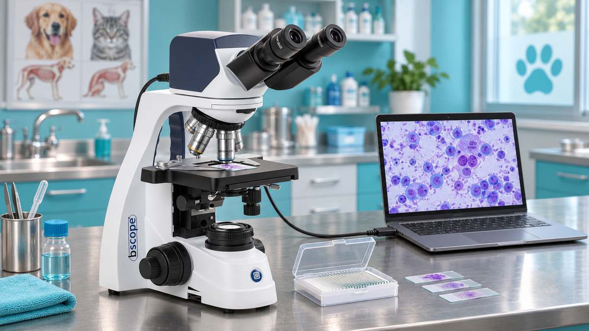
A veterinary microscope is a brightfield biological microscope with 4×, 10×, 40× and 100× oil-immersion objectives. It allows a veterinary practice to examine cytology preparations, blood smears, urine sediment, skin scrapings and faecal samples. For most clinics, a binocular model with good optics and these four objectives is a sensible starting point. If you also want to document cases or teach, consider an imaging option.
This guide explains the applications, the features to compare for your specimens, when to choose a binocular, trinocular or digital configuration, the basic observation sequence and instrument care.
At TiendaMicroscopios, we help you choose a Euromex veterinary microscope according to your usual samples and whether you need to capture images.
At a glance
- Complete cytological assessment requires a good-quality microscope with several objectives, including a 100× oil-immersion objective, as explained in the MSD Veterinary Manual.
- 10× and 40× objectives cover much routine examination of direct faecal preparations and skin scrapings. Blood smears and cytology also call for 100× immersion.
- Choose binocular for routine observation; trinocular or digital when you want to photograph, archive or teach.
- Phase contrast and fluorescence are specialist techniques for particular laboratory applications rather than standard features of a routine brightfield configuration.
- Removing immersion oil after use and keeping the instrument dry help preserve the optics.
Contents
- What is a microscope used for in veterinary practice?
- Which samples are examined and how are they prepared?
- Basic observation sequence
- What to compare when choosing a microscope
- Binocular, trinocular or digital: which configuration?
- What changes for microbiology?
- Care and maintenance
- Our specialists' view
- Where to find your equipment
- Frequently asked questions
- Sources
What is a microscope used for in veterinary practice?
In small-animal practice, microscopy supports the examination of samples during the consultation. A study in the Journal of Veterinary Medical Education highlights the value practitioners place on microscope training for clinical cytology. Applications can be grouped into three settings:
- Clinical practice: skin, ear and mass cytology; blood smears; urine sediment; faecal examination; skin scrapings and hair examinations.
- Laboratory work: reproductive analysis, including semen observation, microbiology, quality control and research.
- Teaching: histology, parasitology and haematology practicals, where sharing the same view with students is useful.
ClinATV describes blood, urine, faecal and cytological tests among common clinical laboratory investigations. Microscopy is part of many of these workflows, alongside other laboratory methods.
Which samples are examined and how are they prepared?
Observation quality depends on specimen preparation as well as the instrument. Even excellent optics cannot compensate for a poorly prepared smear.
Cytology
Adrián Romairone describes common sampling approaches in Diagnóstico Veterinario:
- Impression smear: a slide is pressed against the lesion.
- Swab: useful for skin folds, interdigital spaces and ears.
- Fine-needle sampling: used for swellings and masses.
- Blood smear: supports assessment of blood cells and possible parasitic forms.
- Skin scrapings and hair examinations: used in dermatological investigations.
The Vetiverse recommends preparing cytology slides promptly after sampling, spreading the cells into a thin layer and drying the preparation quickly to preserve morphology. Suitable fixation and staining depend on the sample and procedure. Prepared smears should be protected from temperature extremes.
Direct faecal examination
ClinATV describes direct examination of a faecal suspension to look for structures such as helminth eggs or larvae and motile forms. Its basic sequence is to take part of the sample with a swab, spread it on a slide, mix it with two or three drops of saline, add a coverslip and examine it microscopically.
Blood smear
A paper in Clínica Veterinaria de Pequeños Animales discusses detecting infectious agents on blood smears. Observation requirements vary: some structures can be located at low power, while detailed examination may require the 100× oil-immersion objective. Detection also depends on organism numbers, the sample, the observer's experience and the time devoted to systematic examination.
Basic observation sequence
Ifevet outlines the observation sequence in its blood-smear guide:
- Secure the slide on the stage.
- Start with the 4× objective and use coarse focus to locate the preparation.
- Refine focus with the fine-focus control.
- Move to 10× and refocus.
- Continue with 40× when appropriate.
- For the 100× oil-immersion objective, use a drop of suitable immersion oil.
Low power helps assess the preparation and distribution of material. Higher magnification allows closer examination of morphology. The procedure and objectives used depend on the sample. For blood smears and cytology, a microscope capable of 1000× total magnification with an oil-immersion objective is useful. With a 10× eyepiece, the 100× objective provides 1000× total magnification. 10× and 40× objectives cover much routine faecal and skin-scraping work.
What to compare when choosing a microscope
The MSD Veterinary Manual recommends good-quality optics and a range of objectives, including oil immersion, for complete cytological examination. These are the practical criteria we use when advising a clinic:
| Feature | What to look for | Why it matters |
|---|---|---|
| Objectives | 4×, 10×, 40× and 100× oil immersion | From initial scanning to detailed examination |
| Total magnification | Up to 1000× for cytology and haematology | A 100× objective with a 10× eyepiece gives 1000× |
| Head | Binocular for daily work | Comfort during longer observation sessions |
| Lighting | Adjustable LED illumination | Light intensity can be matched to the specimen |
| Imaging | Trinocular output or a compatible camera | Documentation, consultation and teaching |
| Mechanics | A smoothly moving mechanical stage | Systematic scanning without abrupt movements |
Choose equipment for the samples you actually examine. A configuration that handles your routine work well is more useful than unnecessary specifications.
Binocular, trinocular or digital: which configuration?

Binocular. We recommend the Euromex bScope range for routine clinical observation. The bScope BS.1152-EPL has 10× eyepieces and E-Plan 4×, 10×, 40× and 100× oil-immersion objectives, providing 40× to 1000× total magnification. It includes adjustable 3 W NeoLED illumination, a mechanical stage and coarse and fine focusing.
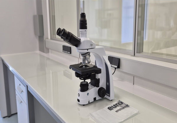
Trinocular. For specimen documentation, shared images and teaching, the Euromex bScope BS.1153-EPL is the trinocular option in our catalogue. Its third tube accepts a camera with a suitable adapter while retaining both eyepieces for observation. Select the camera and adapter for the actual instrument configuration.
Digital. The Euromex bScope BS.1157-EPLi has a 5.1 MP CMOS camera with USB 2.0 connectivity and infinity-corrected E-Plan IOS 4×, 10×, 40× and 100× oil-immersion objectives. You can observe through the eyepieces and capture images on a compatible computer. The microscope does not have an integrated display.

Adding a camera to an existing microscope. If you already use a Euromex bScope, we can help you select a Euromex camera and compatible adapter. Browse our microscope cameras: compatibility and the captured field depend on the head and adapter.
What changes for microbiology?
Routine veterinary microscopy often uses stained preparations and brightfield illumination. Other applications may require a different technique.
- Phase contrast: helps reveal detail in suitable unstained living samples, including movement and structures that staining might alter.
- Fluorescence: visualises fluorescent specimens using appropriate excitation light and filters.
- 100× oil immersion: provides high-magnification examination of stained smears, including bacteria and intracellular parasitic forms.
Check the optical configuration and accessories required for your application. A brightfield bScope does not automatically include phase contrast or fluorescence.
Care and maintenance
The WHO microscope care guide includes practical advice on cleaning and dry storage. After using immersion oil, remove residue with lens tissue and an appropriate optical cleaner. The guide advises against alcohol and xylene for cleaning the lenses.
In warm, humid environments, protect optics from damp and fungal growth. Store the instrument in a dry place and use suitable desiccant storage where recommended. Also:
- Fit the dust cover after use.
- Lower the stage and select the lowest-power objective before storage.
- Clean the immersion objective after every use.
- Check worn objectives, eyepieces and condensers; compatible replacements are available in our accessories and parts section.
Our specialists' view
When a clinic asks us for a microscope, we first ask which samples it examines each week. A quick faecal check, mass cytology and a series of blood smears place different demands on the objectives, head and imaging setup.
We believe the best purchase fits that real workload, with advice from selection through installation and use. We offer personalised guidance and after-sales technical support, and collaborate with the Spanish Microscopy Society.
For most practices, our recommendation is a good-quality biological microscope with up to 1000× magnification. Choose trinocular or compatible camera capture when you need to archive cases or share observations with your team.
The TiendaMicroscopios team
Where to find your equipment
At TiendaMicroscopios, we recommend Euromex bScope microscopes for veterinary applications. We help you choose a binocular, trinocular or digital configuration for your specimens and workflow. Check current commercial and delivery terms when requesting a quotation.

Browse our veterinary microscopes. If you are equipping a classroom, explore our school microscopes and stereo microscopes. Tell us which samples you examine so we can advise you.
Frequently asked questions
What magnification does a veterinary microscope need?
For cytology and blood smears, consider 4×, 10×, 40× and 100× oil-immersion objectives, giving up to 1000× total magnification with 10× eyepieces. 10× and 40× objectives cover much routine faecal and skin-scraping examination.
Can a veterinary microscope also be used for microbiology?
A brightfield biological microscope with oil immersion can be used for stained smears and bacterial observation. Some living-sample or fluorescence applications require a specific configuration, which is not automatically included in a brightfield bScope.
Does my clinic need a microscope camera?
A camera is not essential for direct observation, but it is useful for documentation, comparing images and consulting a specialist. Choose a trinocular instrument or a compatible imaging solution for your existing microscope.
How do I clean the oil-immersion objective?
Remove oil after each use with lens tissue and a suitable optical cleaner. The WHO guide advises against alcohol and xylene for lens cleaning.
Sources
Microscope Use in Clinical Veterinary Practice (Journal of Veterinary Medical Education)
El uso del microscopio en la clínica diaria (II), Diagnóstico Veterinario
El frotis sanguíneo: preparación, identificación y recuento celular, Ifevet
Microscopio óptico o de luz, Manual de microbiología general
Euromex bScope recommendations
Looking for a solution for your centre?
We help you choose the right microscope for your needs.
Discover more
- Choosing a selection results in a full page refresh.